
Contact information
ashley.kamimae-lanning@imm.ox.ac.uk
 https://orcid.org/0000-0002-7516-4881
she/her
Collaborators
-
Verena Korber
Group Leader
-
Marella de Bruijn
Professor of Developmental Haematopoiesis
-

Matthew Nicholls
DPhil Student


Ashley Kamimae-Lanning
PhD
Senior Postdoctoral Researcher
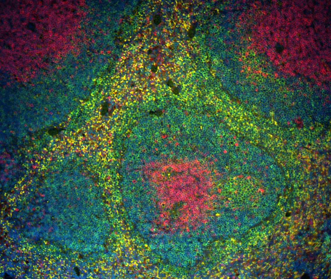
My work in the Patel group is focused on the contribution of endogenous aldehydes to haematopoietic stem cell attrition and squamous cell carcinogenesis in Fanconi anaemia.
I joined the Patel Lab after completing my PhD with Peter Kurre on the in utero programming of haematopoietic stem cells at Oregon Health & Science University. My work in Fanconi anaemia (FA), a rare recessive disease associated with haematopoietic failure, was the first to demonstrate a prenatal defect in the haematopoietic stem & progenitor cell compartment in an animal model. This challenged the conventional paradigm of FA-associated bone marrow failure and highlighted a critical window of opportunity for preventative stem cell therapy.
Protection against aldehyde DNA damage is critical for the haematopoietic system
FA is an inherited disease in which a pathway coordinating the repair of DNA interstrand crosslinks is inactivated. FA is characterised by developmental anomalies, bone marrow failure (often presenting in childhood), and cancer. Most agents known to cause DNA interstrand crosslinks are chemotherapeutic agents such as cisplatin or compounds found in nature such as psoralen, a photoactivated toxin in certain plants; however, the culprits for DNA damage to cells such as haematopoietic stem cells in FA are unknown and are likely to be produced endogenously. Furthermore, mouse models of FA, of which there are now many, have typically had a remarkably mild phenotype compared to that seen in humans. Several years ago, our lab discovered that genetic inactivation of key aldehyde detoxifying enzymes in conjunction with knocking out the FA pathway revealed a phenotype in mice similar to that seen in humans with FA, including spontaneous pancytopenia, bone marrow failure, and leukaemia. This compelling result suggests that endogenous and diet-derived aldehydes such as formaldehyde and acetaldehyde, which can attack DNA to form crosslinks, may be a missing piece of the puzzle in determining what drives the phenotypes seen in FA. We call the dual protective effect of metabolic detoxification and DNA repair "two-tier protection."
Although the catabolism of endogenous aldehydes is critical for preserving the proper function, genomic integrity, and longevity of haematopoietic stem and progenitor cells, it is not known whether this protection must be cell-intrinsic or extrinsic, when it is required, and where the aldehydes are being produced or how far they may be able to travel from the site of production. My project's focus is determining whether detoxification is required within haematopoietic cells or whether other tissues such as liver can provide sufficient detoxificaiton, and examining when in the development of the blood system it is required.
Endogenous DNA damaging agents in squamous cell carcinoma
Patients with FA have an extraordinary, 500- to 700-fold increased risk of developing head and neck squamous cell carcinoma, compared to the general population. However, what drives the development of these cancers in FA is an open question and given that the FA pathway coordinates the complex process of interstrand crosslink repair, we want to examine the role of endogenous formaldehyde. I am investigating what the consequences are of removing two-tier protection from epithelial tissue and whether endogenous aldehydes can initiate squamous cell carcinogenesis.
Recent publications
Metabolite-induced DNA damage drives stochastic stem cell loss and clonal hematopoiesis.
Journal article
Kamimae-Lanning AN. et al, (2026), Cell Stem Cell, 33, 642 - 659.e11
Genotoxic aldehyde stress prematurely ages hematopoietic stem cells in a p53-driven manner.
Journal article
Wang M. et al, (2023), Mol Cell, 83, 2417 - 2433.e7
Two Aldehyde Clearance Systems Are Essential to Prevent Lethal Formaldehyde Accumulation in Mice and Humans.
Journal article
Dingler FA. et al, (2020), Mol Cell, 80, 996 - 1012.e9
Alcohol-derived DNA crosslinks are repaired by two distinct mechanisms.
Journal article
Hodskinson MR. et al, (2020), Nature, 579, 603 - 608
TRAIP is a master regulator of DNA interstrand crosslink repair.
Journal article
Wu RA. et al, (2019), Nature, 567, 267 - 272